
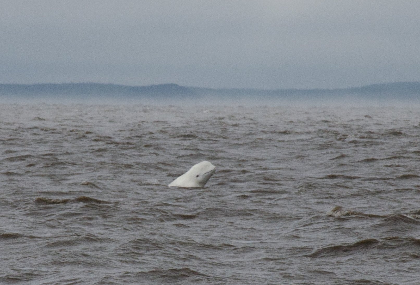
Two female belugas with marked spinal deformities recently washed ashore on the banks of the St. Lawrence River. Although this type of deformation had previously been observed in living animals, no individual had yet been examined up close. These strandings thus enabled the CWHC-Quebec team to investigate the origin of these deformities and assess their potential impact on the health of these two individuals.
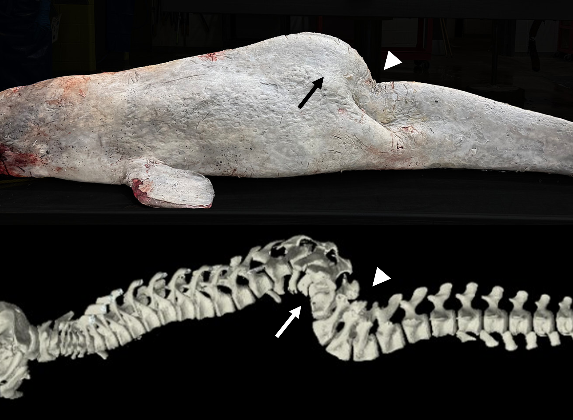
Both belugas were transported to the Faculty of Veterinary Medicine at the Université de Montréal for post-mortem examination and computed tomography (CT scan). Both individuals were emaciated. Imaging studies revealed pronounced spinal curvatures (lordosis, kyphosis) as well as variable scoliosis associated with congenital vertebral anomalies (present at birth) in both cases. The presence of significant degenerative osteoarthritis lesions secondary to the spinal deformities suggests that these bone malformations ultimately contributed to the death of the first beluga, a 17-year-old young adult female. In the second case, a juvenile female of approximately 3 years old, the spinal deformities along with severe parasitic bronchopneumonia were considered as probable co-factors leading to the stranding. In both cases, these vertebral deformities were sufficiently pronounced to have impaired movement, locomotion, and potentially hunting success, contributing to the loss in body condition.
At the population level, a preliminary review of photo-identification data since 2003, conducted by the Groupe de recherche et d’éducation sur les mammifères marins (GREMM), suggests that vertebral deformities are present in fewer than 3% of photographed individuals. Several individuals, some well-known to the marine mammal observation community, have reached adulthood and have been observed for many years. It therefore remains possible for belugas to survive in the St. Lawrence ecosystem despite the presence of vertebral deformities.
Such spinal anomalies have previously been documented in a Scottish population of bottlenose dolphins (Tursiops truncatus); among individuals photographed at or shortly after birth with vertebral deformities (15 individuals representing 7.4% of births over a 23-year period), only three reached adulthood. These deformities, generally presumed to be congenital or associated with trauma occurring at a young age, appeared to progress with growth, presumably ultimately leading to impaired mobility that limited the ability to hunt after weaning or increased the risk of predation (Delynn, 2011; Robinson, 2020). Potential risk factors for congenital vertebral deformities include genetic, nutritional, in utero environmental influences, and chemical exposure (Hersinger, 2009. Acquired causes (occurring after birth) of traumatic origin (collision) or infectious nature are also possible (Bertulli, 2015).
In summary, the two cases presented here merely pave the way for the beginning of investigations aimed at better understanding the underlying causes of these deformities, their potential risk factors, and the impact of these deformities on affected individuals as well as on this endangered population.
This topic will be the subject of a presentation at the annual meeting of the International Association of Aquatic Animal Medicine (IAAAM), which will be held in Montreal from May 10 to 14.
Written by: Émilie L. Couture and Amélia Dalpé
References:
- Bertulli, C. G., Galatius, A., Kinze, C. C., Rasmussen, M. H., Deaville, R., Jepson, P., Vedder, E. J., Sánchez Contreras, G. J., Sabin, R. C., & Watson, A. (2015). Vertebral column deformities in white-beaked dolphins from the eastern North Atlantic. Diseases of Aquatic Organisms, 116(1), 59-67.
- DeLynn, R., Lovewell, G., Wells, R. S., & Early, G. (2011). Congenital scoliosis of a bottlenose dolphin. Journal of wildlife diseases, 47(4), 979-983.
- Hensinger, R. N. (2009). Congenital scoliosis: etiology and associations. Spine, 34(17), 1745-1750.
- Robinson, K. P., Haskins, G. N., Eisfeld-Pierantonio, S. M., Sidiropoulos, T., & Bamford, C. C. (2020). Presenting vertebral deformities in bottlenose dolphin Tursiops truncatus calves from a protected population in northeast Scotland. Diseases of Aquatic Organisms, 140, 103-108.